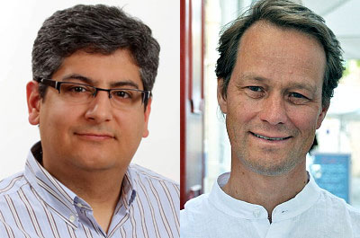
Meza-Zepeda and Myklebost co-author recent Cancer Cell publication on the architecture and evolution of cancer neochromosomes

Leonardo A. Meza-Zepeda and Ola Myklebost in collaboration with David M. Thomas’ group in Australia co-author an article recently published in Cancer Cell (journal impact factor 23.89), entitled “The Architecture and Evolution of Cancer Neochromosomes”.
This article describes for the first time, at single base resolution, the architecture of cancer-associated neochromosomes in well- and dedifferentiated liposarcomas.
Neochromosomes are large marker chromosomes whose origin cannot be determined by conventional chromosome banding. These very large structures contain amplified material from multiple genomic loci, making them different from normal chromosomes. Little is known about the structure of these neochromosomes and the mechanism by which they develop. A number of malignancies have been shown to have this characteristic neochromosome structures, among them well-differentiated/dedifferentiated liposarcoma (WD/ DDLPS). In WD/ DDLPS, neochromosomes have been shown to consistently contain high-level amplifications of chromosome 12q13-q15, with frequent involvement of MDM2 and CDK4 oncogenes. This co-amplification provides a selective benefit for increased survival and proliferation by blocking p53 and RB1, respectively.
By combining chromosome flow sorting and high-throughput sequencing the structure of WD/ DDLPS neochromosomes has been revealed at single nucleotide resolution. Using mathematical modeling, a model for the genesis of neochromosomes has been proposed. This model starts by the formation of episomal structures, stitching together different donor loci in a non-amplifying process suggestive of chromothripsis. These initial structures include the chromosome 12 regions. These double minute structures continue to receive donor sequences through additional chromothripsis cycles. On a second stage, neochromosomes undergo multiple rounds of breakage-fusion-bridge cycles, resulting in amplification and deletion of material, which select for the content of the neochromosome. Under selective pressure, amplified oncogenes are overexpressed, while coamplified passenger genes may be silenced epigenetically. The final stabilizing of the neochromosomes occurs by linearization and telomere capture.
The work is part of a Liddy Shriver Sarcoma Initiative collaborative grant between the Meza-Zepeda’s and Myklebost’s groups in Norway and Thomas’ in Australia. This work has recently been awarded a new extension grant, which also includes Andrew J. Wagner’s group at the Dana-Farber Cancer Institute.
Links:
The Architecture and Evolution of Cancer Neochromosomes
Dale W. Garsed, Owen J. Marshall, Vincent D.A. Corbin, Arthur Hsu, Leon Di Stefano, Jan Schröder, Jason Li, Zhi-Ping Feng, Bo W. Kim, Mark Kowarsky, Ben Lansdell, Ross Brookwell, Ola Myklebost, Leonardo Meza-Zepeda, Andrew J. Holloway, Florence Pedeutour, K.H. Andy Choo, Michael A. Damore, Andrew J. Deans, Anthony T. Papenfuss, David M. Thomas
Cancer Cell, Volume 26, Issue 5, p653-667, 10 November 2014. DOI: http://dx.doi.org/10.1016/j.ccell.2014.09.010
Home page of Leonardo A. Meza-Zepeda's project group - Translational Genomics
Home page of Ola Myklebost's group - Molecular Cancer Biology
