02.06.2022 ESGAR 2022
Abstract ESGAR 2022
Staging of early rectal cancer, the diagnostic performance of MRI to define tumors eligible for local excision.
Ellen Viktil1, Bettina Hanekamp1, Arild Nesbakken2,5, Else Marit Løberg3,5, Ole Helmer Sjo2, Anne Negård4,5 Johann Baptist Dormagen1 and Anselm Schulz1
1Department of Radiology, Oslo, University Hospital Ullevål, Oslo, Norway;
2Department of Gastrointestinal Surgery, Oslo University Hospital Ullevål, Oslo, Norway;
3 Department of Pathology, Oslo University Hospital Ullevål, Oslo, Norway;
4 Department of Radiology, Akershus University Hospital, Lørenskog, Norway;
5 Institute of Clinical Medicine, University of Oslo, Oslo, Norway
Purpose:
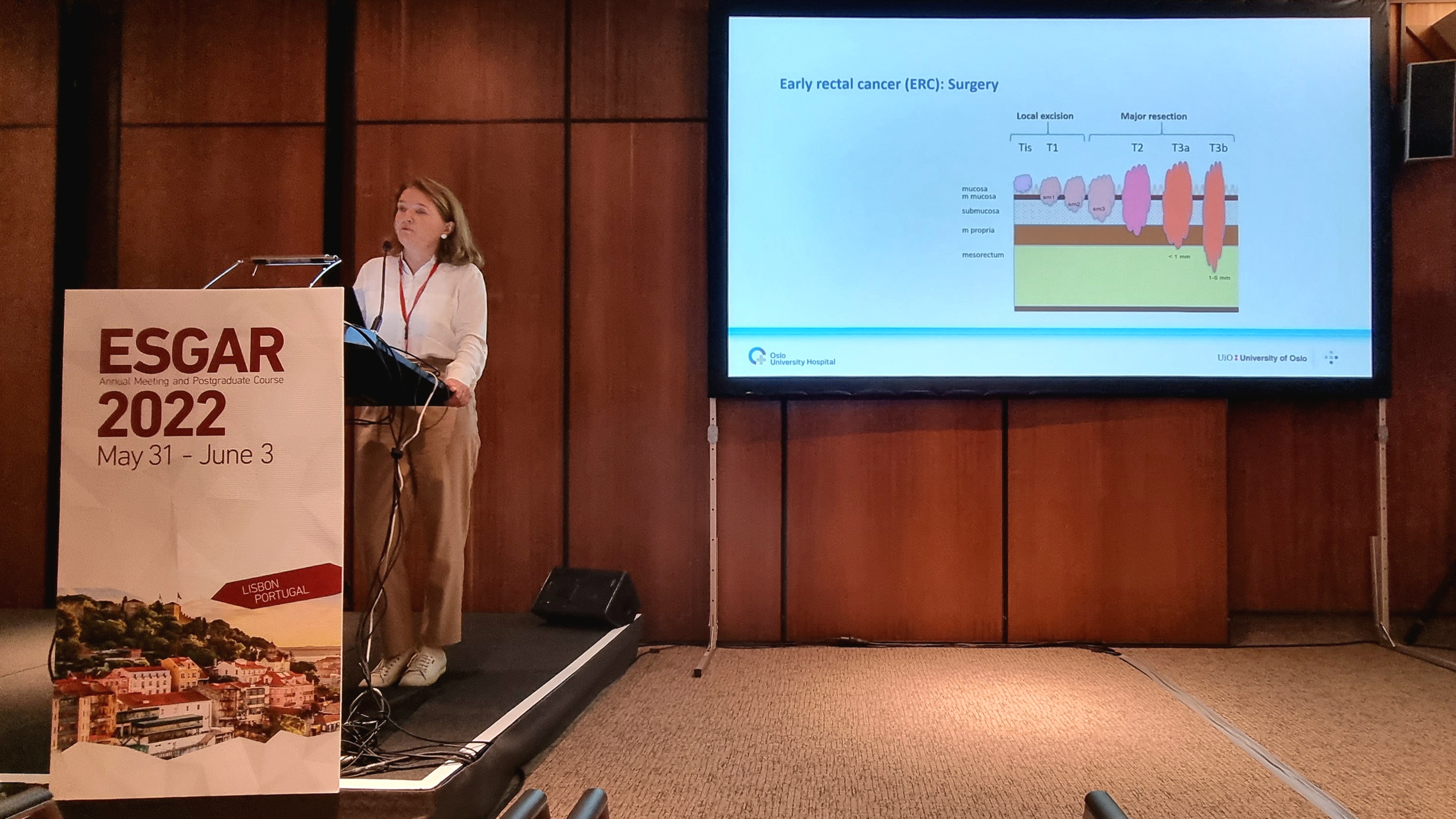
To test the diagnostic performance of MRI for early rectal cancers (ERC) suitable for local excision (LE) when adding a pre-procedural micro-enema and concurrent use of a modified classification system. The micro-enema (Bisacodyl) induces submucosal edema and may hypothetically improve the visualization of tumor depth in submucosa. In addition, we also evaluated the lymph nodes as part of the preoperative staging.
Material and Methods:
In this prospective study, we consecutively included 73 patients with newly diagnosed rectal tumors. Two experienced radiologists independently interpreted the MRI examinations and diagnostic performance was calculated for tumors eligible for LE (Tis-T1sm2, n=43) or as too advanced for LE (T1sm3-T3b, n=30). Each reader registered sensitivity, specificity, PPV and NPV. Inter- and intrareader agreement were assessed by kappa statistics. Lymph node status was retrieved from the clinical MRI reports.
Results:
When using the modified classification system, reader1/reader2 achieved sensitivities, specificities, PPV and NPV of 93%/86%, 90%/83%, 93%/88% and 90%/81%, respectively for identifying tumors eligible for LE. Rates of over-staging of local tumors were 7%/ 14%. Kappa values for the inter-and intrareader agreement were 0.69 and 0.80. For tumors ≤T2, metastatic lymph nodes or nodal tumor deposits were smaller than 3 mm.
Conclusion:
MRI supplemented with a pre-procedure rectal micro-enema and concurrent use of a modified staging system achieved good diagnostic performance to identify tumors suitable for LE. The rate of over-staging of local tumors was comparable to results reported in previous endorectal ultrasound (ERUS) studies. For tumors ≤ T2, metastatic lymph nodes were too small to be characterized by MRI.


