Paper from the Rofstad group highlighted in Cancer Research

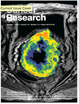
The paper by Tord Hompland et al entitled Interstitial Fluid Pressure and Associated Lymph Node Metastasis Revealed in Tumors by Dynamic Contrast-Enhanced MRI was published and highlighted in the October 1 issue of Cancer Research (journal impact factor 7.856). The cover of the issue shows an illustration from the study, and the paper was highlighted further by a Press Release from the American Association for Cancer Research.
Many malignant solid tumors develop a higher interstitial fluid pressure (IFP) than normal tissues. High IFP in tumors may cause reduced uptake of chemotherapeutic agents, resistance to radiation therapy, and promote metastatic spread. However, measurement of the IFP of tumors poses a challenge because of a paucity of noninvasive imaging strategies. The study by Hompland et al reports the development of a novel method for non-invasive assessment of the IFP of tumors.
Findings were confirmed in cervical cancer patients, where the fluid flow velocity at the tumor surface was higher in patients with pelvic lymph node metastases than in patients without lymph node involvement.
Squamous cell carcinomas of the uterine cervix were subjected to dynamic contrast-enhanced magnetic resonance imaging by using gadolinium diethylene-triamine penta-acetic acid (Gd-DTPA) as contrast agent and an axial T1-weighted spoiled gradient recalled sequence for imaging. The T1-weighted images showed a high-signal-intensity rim in the tumor periphery immediately after the contrast administration, and this rim moved outward with time. The velocity of the rim movement at the tumor surface was associated with tumor interstitial fluid pressure and incidence of lymph node metastases, and may serve as a novel general biomarker of interstitial hypertension-induced tumor aggressiveness.
For details, see article by Hompland and colleagues on page 4899.
Links:
Cancer Research Paper:
Interstitial fluid pressure and associated lymph node metastasis revealed in tumors by dynamic contrast-enhanced MRI.
Hompland T, Ellingsen C, Ovrebø KM, Rofstad EK.
Cancer Res. 2012 Oct 1;72(19):4899-908. doi: 10.1158/0008-5472.CAN-12-0903. PubMed
Press Release, American Association of Cancer Research (ASCR)
