Projects
Cardiac resynchronization therapy
In the project «Non-Invasive Method for Evaluation of Cardiac Resynchronization Therapy (NIME-CRT )» the main aim is to investigate the ability of a simple non-invasive method to evaluate the response cardiac rescynchronization therapy (CRT). Patients who receive CRT have problems with electrical activation of the heart muscle so that some regions of the muscle are electrically activated later than other regions (dyssynchrony). This can be corrected by implanting a pacemaker with electrodes placed in different regions, i.e. CRT. The pacemaker sends an electrical pulse to these electrodes, ensuring that the regions are activated simultaneously. However, about a third of the patients who receive this treatment do not get better. Better methods to evaluate the acute effect of CRT during implantation may aid to locate the optimal pacing sites and pacemaker settings to improve response. It may further be useful during the regular controls of the pacemaker where the effect of CRT on cardiac function can be evaluated with this new method.
Estimation of left ventricular filling pressure
Knowledge of the pressure in the left ventricle while it is filling, is important for making the correct diagnosis of heart failure and monitor treatment. Measurement of this pressure requires a pressure catheter to be inserted into the heart which is an expensive procedure with some associated risks, and hence it is performed in very few patients. The pressure can be estimated risk-free and much less costly by ultrasound imaging by looking at variables related to pressure such as flow velocities and cavity size. We are testing new imaging indices to improve the estimation of filling pressure in ongoing projects.
Blood speckle tracking
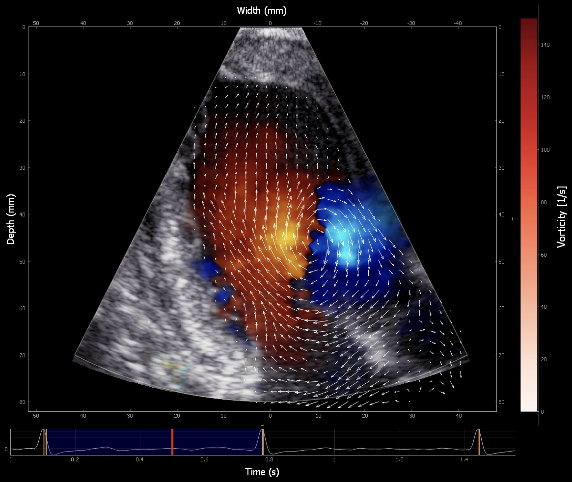
Blood speckle tracking (BST) is a cutting-edge echocardiographic technique used to visualize blood flow in the heart and blood vessels as seen in the image below. BST tracks the speckles created by moving blood cells, similar to myocardial speckle tracking, and uses a very high frame rate to measure high turbulence and flow. Developed by the Department of Circulation and Medical Imaging at the Norwegian University of Science and Technology (NTNU) in Trondheim, this method has been applied in pediatric cardiology, revealing new insights into complex flow patterns. Preliminary data show a strong correlation between non-invasive pressure estimates using BST and invasive pressure measurements in animal models. This novel echocardiographic technology is now available in ultrasound-probes suitable for adults, and we are investigating its feasibility in adult patients with heart failure treated by CRT (NIME-CRT project). Furthermore, we are in the starting phase on projects to investigate how this method can be utilized to evaluate diastolic function and aid cardiac interventions.

Myocardial work
The non-invasive echocardiographic method to assess myocardial work was patented by Kristoffer Russell, Otto Smiseth and Morten Eriksen in our group and it was first published in European Heart Journal in 2012. This method is now commercially available and numerous research groups around the world now apply the method to improve understanding of cardiac function and how the method can be used in clinical evaluation of heart function. Myocardial work is based on the principle of conventional ventricular stroke work which is visualized in a pressure-volume diagram with pressure on the y-axis and volume on the x-axis. One heartbeat then produces a pressure-volume loop, and the loop area represents the work done by the ventricle in one beat. For myocardial work the shortening and lengthening (or so-called strain) of one myocardial segment is plotted on the x-axis instead of volume, and the pressure-strain loop area represents an index of the work done by the segment. Previously myocardial work and stroke work were only possible to obtain when ventricular pressure was measured invasively. However, with the invention of the non-invasive estimated pressure trace, it became possible to obtain myocardial work non-invasively. We are currently combining the estimated pressure trace with measured volume trace from 3D echocardiography and validate this non-invasive method and its applicability in different conditions. We further develop and utilize these non-invasive methods in different studies.
Transcatheter aortic valve implantation (TAVI)
Aortic stenosis is one of the most prevalent heart disorders and has traditionally been treated with open-heart surgery replacing the diseased valve. Over the last decade, there has been an exponential growth in the number of aortic stenosis patients treated with transcatheter aortic valve implantation (TAVI), which compared to surgery is a far less invasive procedure and provides good results. There are, however, several unanswered questions related to TAVI. We are currently exploring why there is discordance between echocardiograpic and invasive pressure gradients in TAVI patiens. This discrepancy is important as it may lead to an inaccurate assesment of valvular function. Another concern is the rapidly increasing number of TAVI patients in need of a second valve intervention. We are planning studies on both the feasibility for such procedure and how to optimize hemodynamics with a second valve.
