Services

We provide services within a range of imaging techniques, such as confocal, live-cell, and various superresolution imaging techniques. We routinely train users in microscopy, enabling them to independently operate our microscopes, while we also offer microscopy as a service. Contact us if you would like to make an appointment for training. We also offer to have meetings with users to discuss imaging needs and options. Contact us by mail for further information and/or protocols for sample preparation.
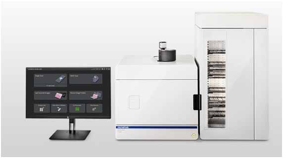
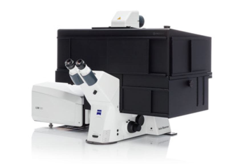
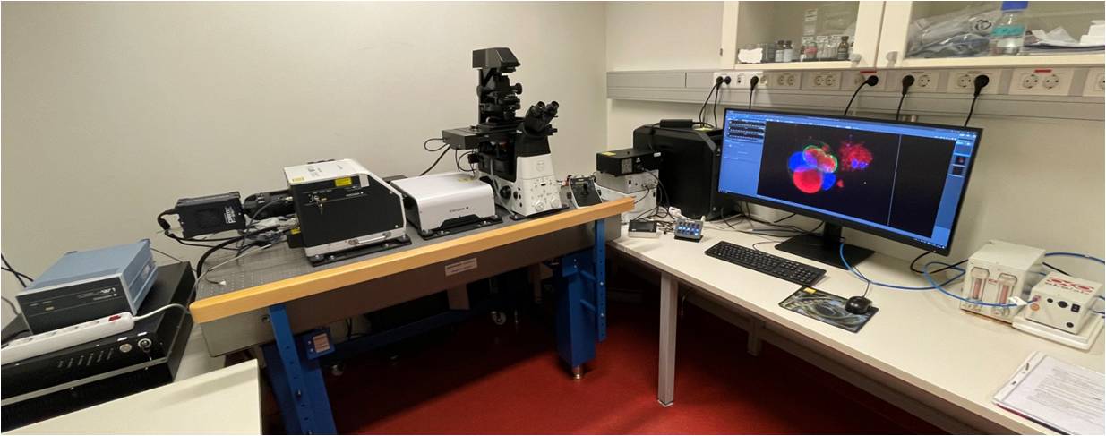
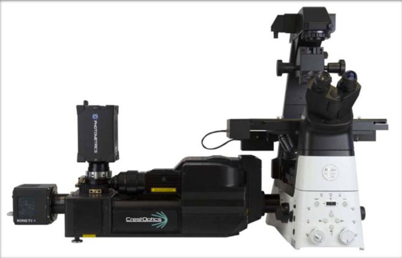
 Price List

Equipment

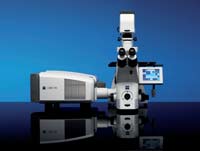
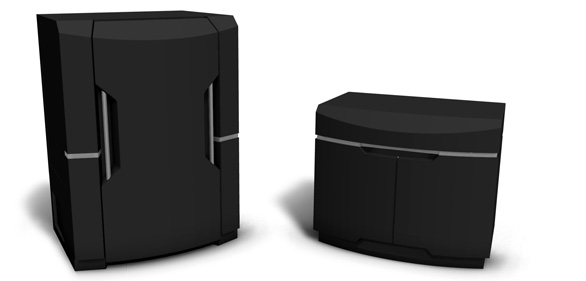
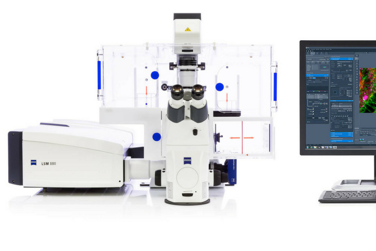
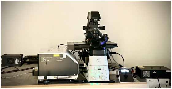
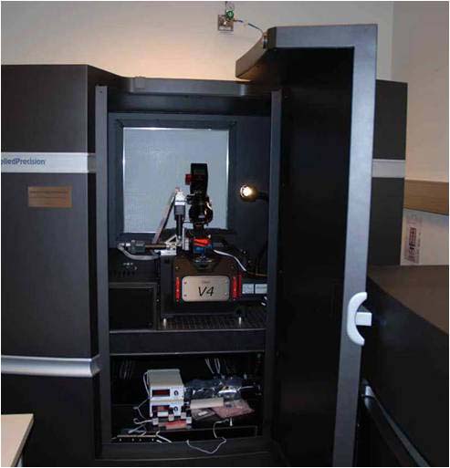
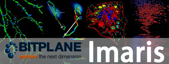
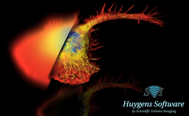
The Core Facility for Advanced Light Microscopy, Montebello holds a Zeiss LSM 880 Airyscan FAST, a Zeiss LSM 980 Airyscan, and a Zeiss LSM 780 confocal microscopes, a DeltaVision OMX V4 Blaze super-resolution microscope, a Nikon Ti2-E microscope with a Yokogawa CSU-W1-SoRa spinning disk and a Nikon Ti2-E with a CrestOptics V3 spinning disk, an Olympus VS200 slide scanner, as well as software licences for Imaris and Huygens.


LSM 980 Airyscan 2

LSM980 is an inverted, confocal microscope with Airyscan2 for superresolution, and is equipped for live-cell imaging including a Definite Focus system. It comprises six laser lines (405, 458, 488, 514, 561,  and 633 nm). Available objectives: 5x, 20x NA 0.8 DIC II (Plan-Apochromat), 25x NA 0.8 (LD LCI Plan-Apochromat), 40x NA 1.2 Water Imm DIC III (C-Apochromat), 63x NA 1.4 oil DIC III (Plan-Apochromat). The system includes an AI sample finder, a Colibri 7-color LED, and an Axiocam 702 camera for widefield imaging.

Available imaging techniques:

  • Multicolor fluorescence confocal imaging
  • Two-and three-Dimentional Imaging (Z-stack)
  • Time Series (Live Imaging) in temperature and CO2 controlled environment
  • Airyscan superresolution including SR, SR-4Y and SR-8Y multiplex modes.
  • Widefield imaging
  • ZEN Connect

LSM 880 Airyscan FAST

LSM 880 Airyscan FAST is a confocal microscope equipped with an Airyscan detector and FAST options. The airyscan detector gives possibilities for superresolution imaging with a resolution upto 1,7 times that of conventional confocal imaging. The airyscan detector also provides high sensitivity and can be combined with the FAST beam shaper to give fast and gentle live-cell imaging.

Laser lines: 405, 458, 488, 514, 561,  and 633 nm. Available objectives: 10x NA 0.45 DIC II (Plan-Apochromat), 20x NA 0.8 DIC II (Plan-Apochromat), 25x NA 0.8 (LD LCI Plan-Apochromat), 40x NA 1.2 Water Imm DIC III (C-Apochromat), 63x NA 1.4 oil DIC III (Plan-Apochromat). Definite focus 2 system. Temperature and CO2 controlled chamber.

Available imaging techniques:

  • Multicolor fluorescence confocal imaging
  • Two and Three-Dimensional Imaging (Z-stack)
  • Time Series (Live Imaging)
  • FRAP, FRET, Photoactivation
  • Airyscan superresolution
  • Airyscan FAST imaging

Nikon ECLIPSE Ti2-E Yokogawa CSU-W1-SoRa spinning disk

The Nikon SoRa system is a Nikon ECLIPSE Ti2-E microscope equipped with a CSU-W1 spinning disk confocal unit (50 um pinhole size) and a SoRa microlensing disk for superresolution.

The system is equipped with two Prime BSI cameras (back-illuminated sCMOS) for sequential or two-color simultanous imaging.

The system is suitable for fixed as well as live sample imaging, and is equipped with a Okolab stage top incubator for temperature and CO2 control.

SoRa superresolution imaging is possible with 60x or 100x objective and can give a resolution of 120 nm in XY.

Available objectives:

  • 4x
  • 10x Air
  • 20x CorrectionCollar for upto 1,2 mm coverglass thickness
  • 25x Silicone immersion
  • 40x Air
  • 60xWI with automatic water immersion dispenser
  • 100x Oil immersion
  • 100x Silicone immersion 

Nikon ECLIPSE Ti2-E CrestOptics X-Light V3 spinning disk

The Nikon Crest system is a Nikon ECLIPSE Ti2-E inverted microscope equipped with the CrestOptics X-Light V3 spinning disk confocal module.

The Nikon Crest spinning disk microscope is suitable for imaging live cells, fixed samples, thick tissue samples, as well as organoids. This instrument has a large field of view (25 x 25 mm) and uniform illumination resulting in seamless stitching of large images.

7 laser lines allows imaging of the most common fluorophores (DAPI, FITC, TRITC, Cy5, Cy7, CFP, YFP) in either cofocal mode or in widefield mode.

In addition, the dual camera setup and Z piezo device give fast imaging of multiple channels and z-stacks with nanometer precision.

Microscope features:

  • Motorized XY stage
  • Z piezo device
  • Stage-top incubator and XL-cage with CO2 and temperature control
  • Dual camera imaging (Back-illuminated sCMOS cameras, Photometrics Kinetix)
  • Lumencor Celesta multi-line laser (405, 446, 477, 518, 546, 638, 749 nm)
  • CrestOptics X-Light V3 spinning disk (50 um pinhole)
  • Objectives:

CFI Plan Apo l D 10X NA 0.45 (Air)

CFI Plan Apo l D 20X NA 0.8 (Air)

CFI Plan Apo l D 40XC NA 0.95 (Air)

CFI Plan Apo l S 40XC Sil NA 1.25 (Silicone)

CFI Plan Apo lIR 60X NA 1.27 (Water)

CFI Plan Apo l D 60X Oil NA 1.42 (Oil)

CFI Plan Apo l D 100X Oil NA 1.45 (Oil) 

  • Tube lens changer (1.5x extra magnification)
  • Anti-vibration table
  • NIS Elements AR software

OMX

The DeltaVision OMX V4 Blaze 3D-SIM microscope is an advanced multimode, super-resolution microscope system.

Imaging options:

  • SIM: structured illumination microscopy for multicolor 3D super-resolution imaging. Gives a resolution down to 110 nm lateral and 330 nm axial.
  • Localization microscopy (STORM): 2D super-resolution imaging with a resolution down to 20 nm.
  • Conventional mode: ultra fast widefield imaging, with temperature and CO2 control for live cell imaging.
  • TIRF: Total Internal Reflection Fluoresence, for enhanced signal-to-noise imaging of the surface of a cell/specimen.
  • PhotoKinetics/Photoactivation imaging system for photoactivation, FRAP, and FRET imaging methods.

Other features of the OMX: 6-color solid state illumination and 6 lasers (405 nm, 445 nm, 488 nm, 514 nm, 568 nm, and 642 nm), UltimateFocus Hardware Autofocus System, three high speed sCMOS cameras. Objectives: 60X NA 1.42 oil PLAPON6 PSF, 60X NA 1.49 TIRF, and 60X NA 1.30 Sil. 

Protocol for new users of SIM: OMX-SIM

Olympus VS200 Slide scanner

The SLIDEVIEW VS200 digital slide scanner is equipped with a loader for upto 210 slides, robotics for automatic loading of slides into the scanner, and light sources and cameras for both brightfield and fluorescence image scanning. It has automatic sample detection,  Autofocus system,  a 2x objective for overview-scans, and 4x, 10x, 20x, 40x and 60x objectives for high quality, quantitative detailed imaging. The system can automatically scan overview and detailed images of samples mounted on glass-slides (standard 26x76mm, or larger).

Imaging Techniques:

  • Brightfield imaging of tissue/tissue sections stained with HE, HRP-immunostain, or others.
  • Fluoresence imaging of upto four colors (blue, green, red and farred).
  • 3D/Z-stack imaging
  • Multibatch scanning of slides

Image analysis

Imaris - Imaris (Bit-Plane) licence version 9.0.2, which includes algoritms for Visualization, Segmentation, Classification and Statistical Analysis of Surfaces, Spots and Contours, for Tracking Objects, and for Colocalization Measurements.

Huygens- Huygens Essential, version 14.06, image processing software for deconvolution, visualization and analysis.