
Correlative Light Electron Microscopy (CLEM)
Light microscopy (LM) such as fluorescence microscopy offers the advantage of providing wide-field imaging of dynamic processes in living samples. However, its resolution is limited, and importantly fluorescence microscopy only allows researchers to visualize the specific tags added to the samples and it does not provide structural information to be certain of the localization of this tag to a specific organelle. In contrast, electron microscopy (EM) delivers much higher resolution images, enabling detailed examination of ultrastructural features. However, EM has a more limited field of view and can not be used for live imaging.
The correlative light-electron microscopy (CLEM) technique effectively combines these two methods to harness their strengths. In CLEM, fluorescently marked structures or tagged proteins are tracked in living cells with LM, allowing for real-time observation of their behavior. The fluorescent patterns identified through LM can then be confirmed at the ultrastructural level. This integration allows researchers to identify cellular structures and processes of interest in whole-cell images captured by LM and subsequently conduct detailed analyses with EM, which may include advanced techniques such as immuno-electron microscopy and electron tomography.

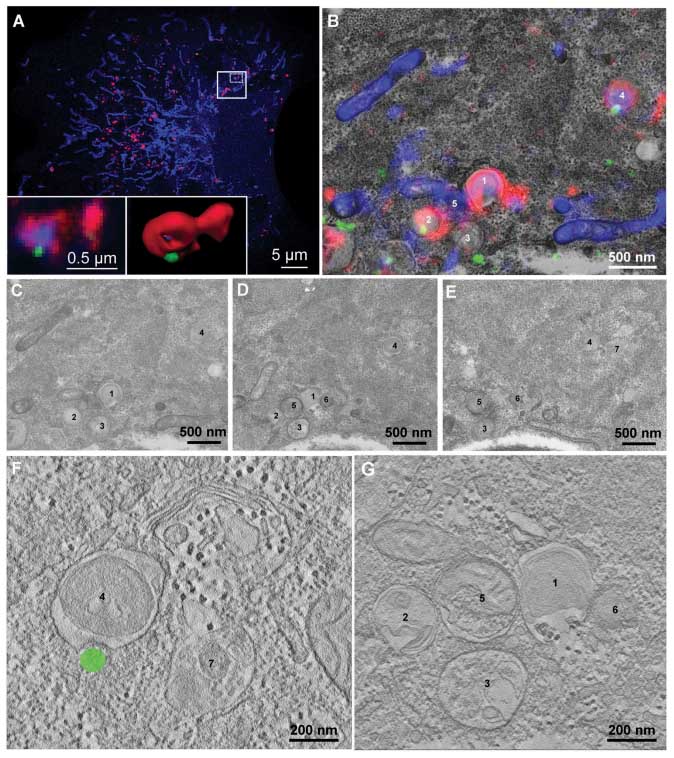
Vietri et al 2014, Nature Letters, CLEM |
|
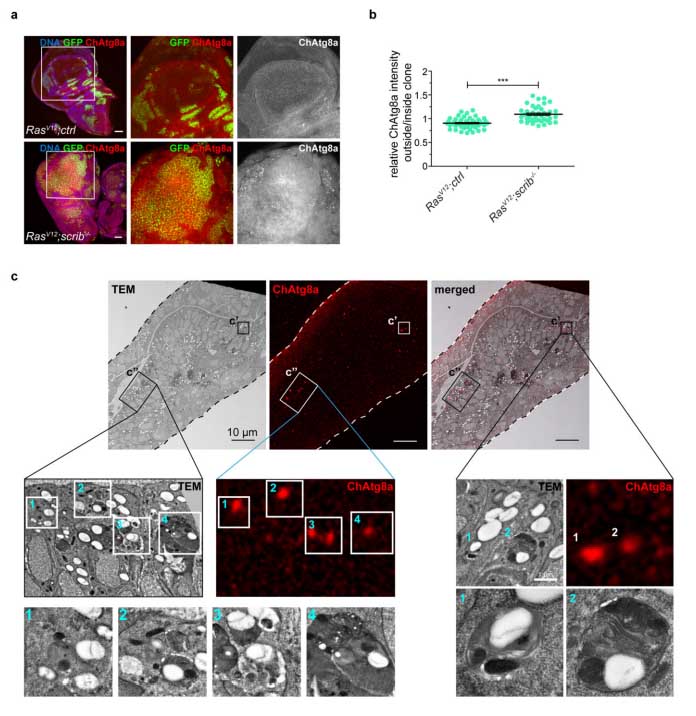
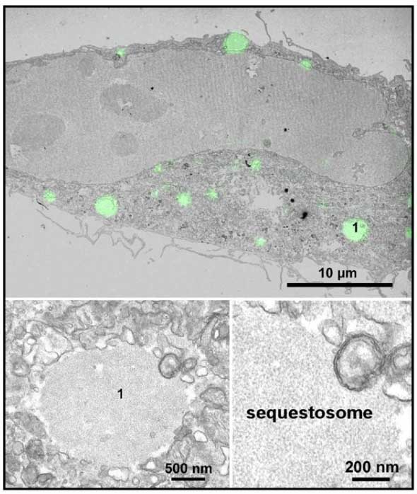
Katheder et al 2017 Nature, CLEM |
|
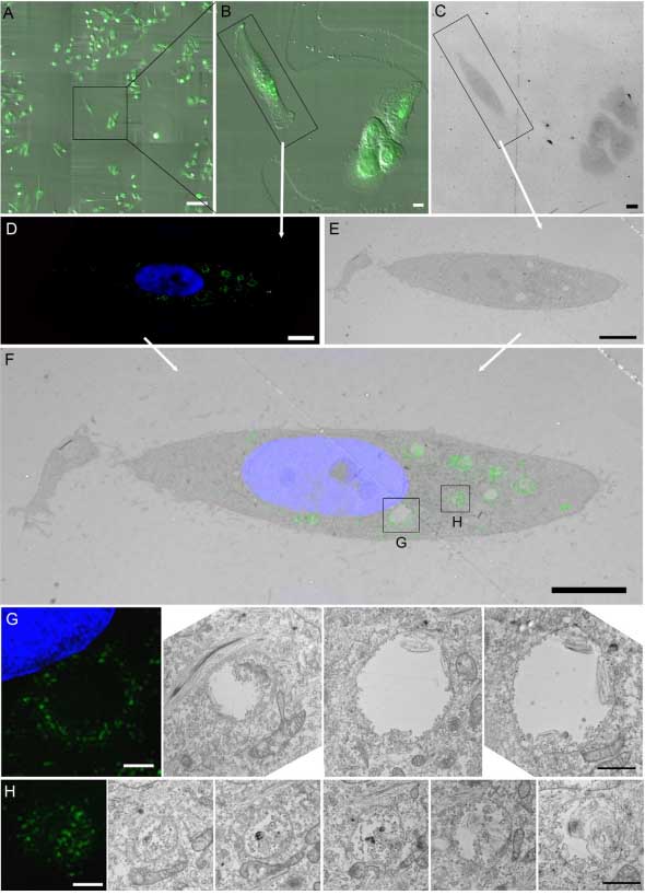
Wegner et al 2014, Methods in Enzymology, CLEM |