E3 ubiquitin ligases in intercellular communication and carcinogenesis

The responsible for the preclinical work on E3 ubiquitin ligases is Dr Edward Leithe, who leads the "Cell Signaling" project group.

Objectives:

1) Define the role of selected E3 ubiquitin ligases in colorectal cancer pathogenesis and their potential as biomarkers.

2) Identify and functionally characterize E3 ubiquitin ligases that regulate intercellular communication via gap junctions in normal and cancerous cells.

Background:

Ubiquitination is a posttranslational modification where ubiquitin, a small globular protein, is covalently conjugated to a target protein. The reaction is a multistep process involving three classes of enzymes, of which the E3 ubiquitin ligases provide specificity to the system by selecting the substrate protein. The E3 ubiquitin ligases interact with and ubiquitinate protein substrates in a temporally and spatially regulated manner, and these processes are frequently deregulated in human cancers. Several E3 ubiquitin ligases represent attractive drug target candidates. Gap junctions consist of intercellular channels that provide for direct cell-to-cell movement of signaling molecules and ionic currents. Gap junctions have important roles in maintaining cellular homeostasis, and loss of these structures during cancer pathogenesis may contribute to increased cell growth and radio- and chemotherapy resistance.

Precision medicine of colorectal cancer

Objectives: (i) develop a platform for tumor organoid culturing and ex vivo drug sensitivity testing to model cancer treatment in a personalized manner; and (ii) identify new treatment options as well as biomarkers and mechanisms of treatment response and resistance by integrated pharmacogenomics analyses.

Background: Almost 900,000 patients die from colorectal cancer every year, and the main cause of death is metastasis to the liver. Overall survival among patients with metastatic colorectal cancer has increased to almost 3 years with currently available therapies. Recent advances hold promise to further improve treatment efficacy by biologically-based patient stratification, but the number of available drugs is low compared to other major cancer types. Each “actionable” molecular marker is found in only 1-5% of the cancers, and the overall clinical benefit is modest (with the notable exception of long-lasting responses to immune checkpoint inhibitors in MSI cancers) (Sveen, Kopetz and Lothe, Nat Rev Clin Oncol 2020). Drug sensitivity testing offers a complimentary approach to personalized treatment.

Immune check point inhibition in microsatellite unstable CRC: we discovered that the microsatellite instability hypermutation phenotype is present in a subgroup of sporadic CRC and that it is a marker of good prognosis (Lothe et al., Cancer Research 1993; cited > 800 times). We also found that MSI is present in subgroups of other solid tumors (Peltomaki et al., Cancer Research 1993; cited > 600 times). MSI was in 2017 approved by FDA as the first pan-cancer biomarker, predicting benefit from immune checkpoint blockade in metastatic cancers. Despite the strong effect of this therapy on clinical care, only half of the MSI positive metastatic cancers respond. We have identified a resistance mechanism and negative predictive marker (Sveen et al., Genome Med, 2017).

Outcomes: preclinical resources and underlying research. We have established large preclinical pharmacogenomics data resources including (i) >100 conventional colorectal cancer cell lines screened for sensitivity to ~500 drugs and analyzed for gene expression and mutations; and (ii) a living biobank of >300 tumor organoids of liver metastases (>1 metastatic lesion from 65% of 150 patients), all screened for sensitivity to 40-73 selected drugs and analyzed for gene expression and mutations.

We developed CMScaller, a computational tool to translate the consensus molecular subtypes to pre-clinical models, and used our cell line resource to identify the specific drug vulnerabilities of each subtype (Sveen et al. Clin Cancer Res 2018, Eide et al. Sci Rep 2017, Berg et al., Mol Cancer 2017). We published the first large study of inter-metastatic heterogeneity of drug sensitivities in tumor organoids of colorectal cancer, and showed that organoids can model development of chemoresistance in a personalized manner (Bruun et al., Clin Cancer Res 2020 and Kryeziu et al., J Transl Med 2021). We have used this personalized treatment approach to successfully guide the rechallenge with chemotherapy in a patient who had exhausted all treatment options (in Norwegian). Ongoing analyses of the complete living biobank confirm that organoids are good personal cancer models also on the molecular level, and we develop models to predict new drug vulnerabilities based on histopathological and molecular subtypes. We have shown a biological rationale for repurposing of PARP-inhibitors to colorectal cancers with wild-type TP53 activity (Smeby et al., EBioMed 2020), and clinical translation is pursued in the EVIDENT trial.

Outcomes: functional oncology trial
Using our living biobank as a reference for personalized treatment nominations, we are currently conducting a functional oncology trial of metastatic colorectal cancer. EVIDENT (ex vivo drug sensitivity testing; ClinicalTrials.gov Identifier: NCT05725200) is a prospective intervention study of tumor organoid-guided treatment in the context of tumor heterogeneity. The trial combines standard precision cancer medicine approaches and analyses of “actionable” biomarkers with drug sensitivity testing of both chemotherapies and experimental cancer drugs. The trial evaluates the potential to translate tumor organoids from research tools to treatment decision tools.

Tumor heterogeneity of colorectal cancer

We aim to uncover the clinical consequences of spatio-temporal tumor heterogeneity of primary and metastatic colorectal cancers. We use “multi-omics” technologies and data integration to map molecular tumor profiles in the context of tumor heterogeneity. Spatial heterogeneity is analyzed among multiregional samples of each tumor and/or multiple metastatic lesions from each patient. Metastatic evolution is analyzed by comparisons of patient-matched primary tumors and metastases.

Biomarkers of colorectal cancer

Colorectal cancer is the third most common type of cancer in the world, and the incidence is increasing both among young and elderly individuals. Chemotherapy after surgery of locoregional colorectal cancers improves the survival rate by approximately 15%. However, not all patients benefit from chemotherapy, and both over- and under-treatment is frequent when following the current treatment selection criteria. Personalized approaches to adjuvant chemotherapy can improve the risk-benefit ratio. For this purpose, we identify biomarkers to predict patient prognosis and treatment benefit. We have identified prognostic gene expression-based signatures (Ågesen et al., Gut 2012 and Sveen et al., Clin Cancer Res 2012) and protein markers (Bruun et al., Clin Cancer Res). Further improvement to the diagnostics of colorectal cancer is likely to be specific for smaller subgroups of patients, and be based on biomarkers of low prevalence and/or a combination of several biomarkers (Smeby et al., Ann Oncol 2018). We contribute to international multi-center studies analyzing thousands of patients to identify new and robust biomarkers, including POLE mutations causing “ultramutated” tumors (Domingo et al., Lancet Gastroenterol & Hepatol 2016), markers of the tumor microenviroment (Dienstmann et al., Ann Oncol 2017 and 2019; Glaire et al., Brit J Ca 2019) and protein expression markers (Bergsland et al., ESMO Open 2020).

Testicular cancer: Tumour stem cells, etiology and effects after chemotherapy

Tumour stem cells and germ cell cancer


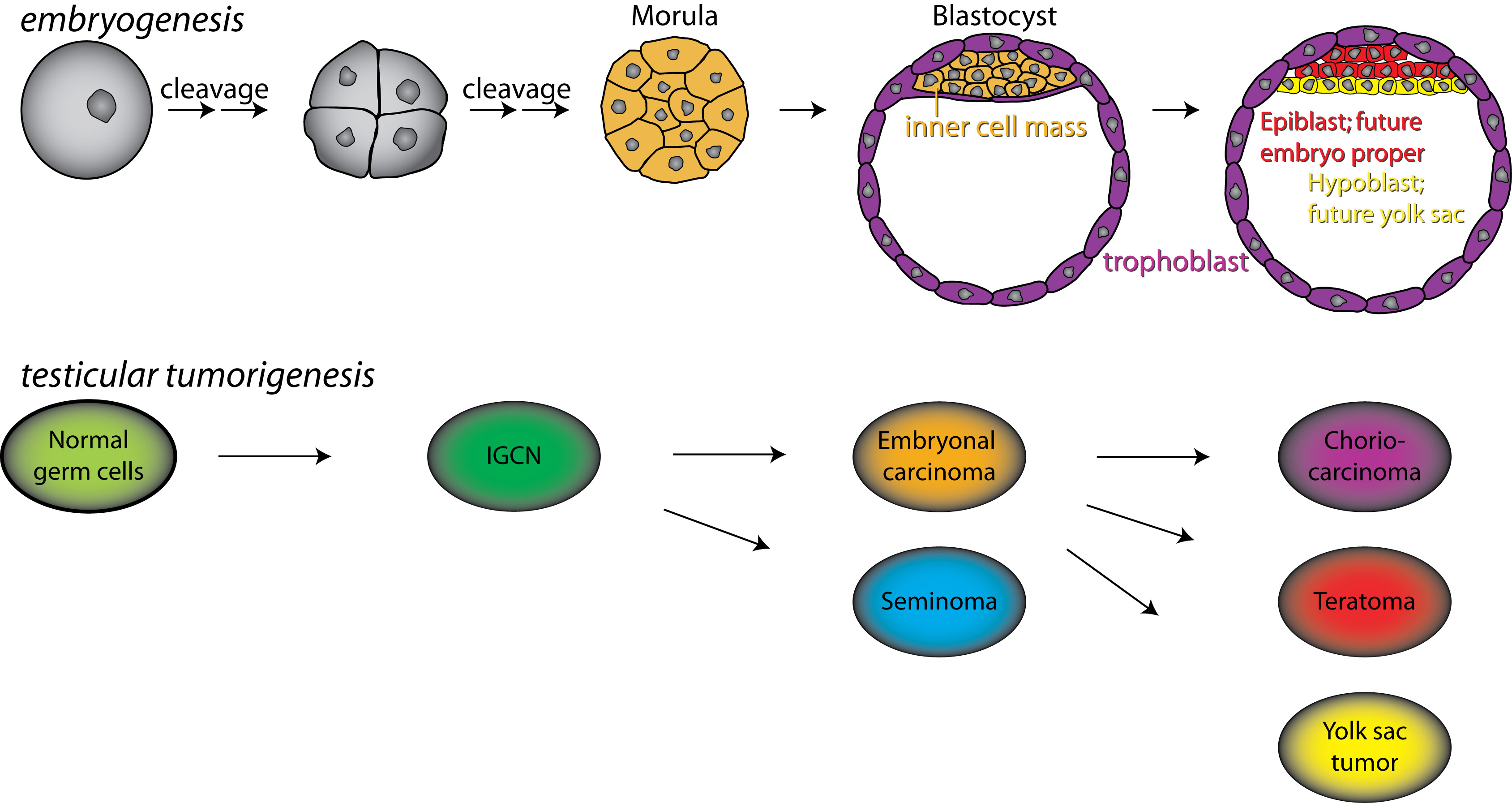
(A) Comparisons of embryonal carcinomas and embyronic stem cells (Figure and text below).
(B) Genetic risk and long-term effects of treatment as assessed from genotypes in DNA repair genes in germ cell tumour patients.


Embryonal carcinoma (EC) cells, found in certain testicular germ cell tumours (TGCTs), represent a striking paradigm for malignant cells with stemness properties. Their pluripotent state allows them to self-renew, as well as differentiate, to give a disorganized array of cell and tissue types. EC cells are morphologically similar to embryonic stem (ES) cells, and the two also share cell surface markers and overall gene expression programmes.


Overall, this imply that ES cells can be considered the non-malignant counterpart of EC cells, providing the rare situation where the "normal" counterpart of a cancer cell with stemness properties is readily accessible, and can be observed through its progression to the malignant state.


By comparing gene expression of EC and ES cells across the genome at high resolution, we aim to gain insight into the role of stem cells in the development and progression of TGCTs, in particular, and cancer in general.


Genetic variation among testicular cancer patients; predisposition, chemotherapy response and long term toxicity

Testicular germ cell tumors (TGCTs) are the most common tumors among young men, aged 15-45 years. The TGCTs are histologically classified as seminomas and nonseminomas, both believed to originate from a common precursor known as intratubular germ cell neoplasia.


Fortunately, these tumors respond well to cisplatin. More than 95% of all patients and above 80% of patients with metastasis are cured using platinum based chemotherapy. However, some patients are troubled by side effects and long term toxicities of their treatment and also some tumors do not respond to treatment. Based on previous studies of TGCTs, we have selected a number of candidate single nucleotide polymorphisms (SNPs) to be profiled among the TGCT patient population.


We aim to discover if these genetic variations could be involved in and/or contribute to predisposition and development of TGCTs, influence the tumors' response (cure or relapse) and the patients' response (long term side effects/toxicities) to treatment. We hope to identify SNPs that may be applied for more individualized diagnostics and therapy selection, and prevention of TGCT.


The research project is run jointly between the Molecular Genetics Group and the Genome Biology Group.

Adoption of an orphan: Malignant peripheral nerve sheath tumors

Background
Malignant peripheral nerve sheath tumors (MPNST) are rare soft tissue cancers that arise in neuroectodermal cells in the peripheral nervous system. About half of the patients carry the additional burden of Neurofibromatosis type 1 (NF1), which is a hereditary genetic disease that significantly increases risk of developing MPNST, as compared to the general population. Although MPNST is rare, there is a great need to improve the current treatment, as most patients are young adults and the survival prognosis is poor. The current treatment of MPNST patients includes surgery, chemotherapy and radiotherapy, however, there is little evidence to support the benefit of the treatment options beyond surgery.

Goal
We aim to create and transfer biological discoveries into improved management of patients with MPNST.

Ongoing research
In a European multicentre study led by Prof Lothe we have in recent years identified prognostic biomarkers that can be used for stratification of high risk patients who could benefit from additional treatment after surgery and low risk patients who might be spared from the burdens of aggressive treatment. Current research is focusing on identification of predictive markers that can guide therapy choice for patients. We also investigate preclinical models aiming to identify novel drug sensitivities and synergistic drug combinations. Multilevel molecular profiling is performed on fresh frozen tumor samples. Clinical translation is performed in collaboration with Dept of Oncology, OUH.
 
Clinical trial with Simon’s two-stage design:
A phase II, single arm interventional trial of pembrolizumab (anti-PD-1 antibody) in patients with metastatic or locally advanced/non-resectable MPNST. ClinicalTrials.gov (NCT02691026). Translational substudy ongoing.

Collaborators

Oslo University Hospital
Assoc. Prof. Bodil Bjerkehagen, Dept of Pathology
Dr Kjetil Boye, Dept of Oncology
Dr. Tormod K Guren, Trial Unit, Dept of Oncology
Prof. Sigbjørn Smeland, Division of Cancer Medicine

European Centres
Prof. Fredrik Mertens, Skåne University Hospital, Lund, Sweden
Dr. Emanuela Palmerini and Prof. Piero Picci, Istituti Ortopedici Rizzoli, Bologna, Italy
Dr. Eva van den Berg, University Medical Centre, Groningen, the Netherlands