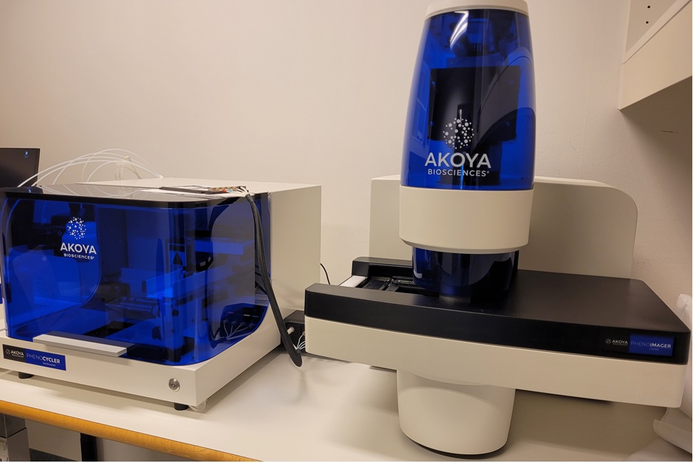
Akoya PhenoCycler Fusion 2.0

The PhenoCycler Fusion system from Akoya enables multiplex spatial phenotyping of tissue sections at single-cell and sub-cellular resolution. You can do spatial mapping of both protein and RNA biomarkers in paraffin-embedded or fresh-frozen tissues.
The instrument uses high quality optics to acquire images with 10 - 40x magnification. Typically, the 20x magnification gives a resolution of 0.5µm/pixel, while 40x enables a resolution of 0.25um/pixel.
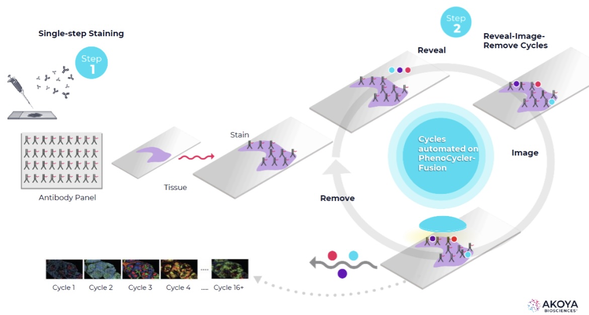
The PhenoCycler Technology combines automated fluidics with high-speed imaging (see illustration below). There is one staining step using barcoded antibodies, following repetitive binding and unbinding of the corresponding fluorescent reporters.

The PhenoImager can also be used for acquiring images on tissue stained with the Opal dyes.
Microscope features:
- Spatial mapping of protein & RNA biomarkers in tissue sections
- High-plex spatial mapping (+60 biomarkers) of two tissues simultaneously
- Cellular & sub-cellular resolution
- PhenoCycler Fusion technology (barcoded antibodies + corresponding fluorescent reporters)
- PhenoImager: Acquisition of tissue stained with Opal dyes
- FFPE or fresh-frozen tissue sections
- Acquisition of HE-stained tissue
Contact:
 |
Anna Lång, PhD Telephone: +47 23013915 E-mail: a.u.lang@medisin.uio.no |
