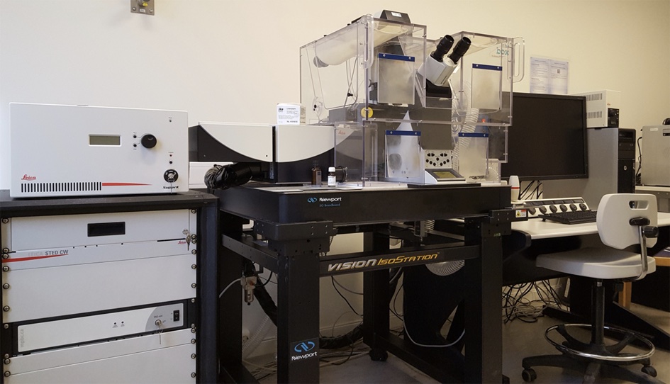
Gated STimulated Emission Depletion (gSTED) Microscopy

gSTED microscopy is a further development of conventional confocal microscopy and acquires images by point scanning over the sample, generating images pixel by pixel. Adding a second laser beam, depletion beam, to the normal excitation laser allows for specific emission of fluorescent dyes within very small 20-80 nm diameter regions in the sample. This achieves sub-diffraction xy resolution, also known as super-resolution.
Technically this works by surrounding the diffraction-limited excitation laser spot with a doughnut shaped “sheath” of depletion light, with a central zero-intensity spot in the focal plane. During STED microscopy, a tunable white laser creates an ordinary, diffraction-limited spot of excited molecules in the sample, while the STED laser acts on the excited dye molecules, quenching them to the ground state. This phenomenon is known as STimulated Emission Depletion (STED).
The net effect of the STED laser is that the excited dye molecules affected by it do not fluoresce. By the spatial arrangement of the STED laser beam in to a doughnut-shape, surrounding the excitation spot, it is possible to collect only the fluorescence from dye molecules at its very center, thus breaking the diffraction barrier. In addition, the Leica TCS SP8 system uses a time-gated detector, which further improves image resolution and signal to noise ratios in both STED and conventional confocal mode.
Our system is also equipped with a CO2 and temperature-controlled incubation chamber for live-cell super-resolution imaging. For single- and two-color STED, we provide compatible fluorescently labeled secondary antibodies, high-precision cover glasses, and STED-compatible mounting medium, if required.
Microscope features:
- Tunable white light excitation laser, with up to 8 simultaneous laser lines (470-700 nm)
- UV laser (405 nm)
- 592 nm STED laser
- 4 adjustable photodetectors (2 time-gated HyDs and 2 PMTs)
- 20x 0.75 NA HC PL APO CS2 immersion objective, correction collar set to Oil
- 20X 0.70 NA HC PL APO air objective
- 40X 1.10 NA HC PL APO CS2 continuous-film objective with water immersion micro dispenser, DIC
- 40X 1.3 NA HC PL APO CS2 oil immersion objective, DIC
- 100X 1.4 NA HCX PL APO oil immersion objective (STED), DIC
- Live-cell incubation chamber with CO2 and temperature-control
Further reading:
STED microscopy description from Stefan Hell’s (inventor of STED) lab:
http://www3.mpibpc.mpg.de/groups/hell/STED.htm
STED literature references on MicroscopyU:
http://www.microscopyu.com/references/superresolution/sted.html
Quick Guide to STED Sample Preparation
https://www.sigmaaldrich.com/content/dam/sigma-aldrich/docs/Sigma-Aldrich/General_Information/1/sted-sample-preparation-guide.pdf
Contact:
 |
Anna Lång, PhD Telephone: +47 23013915 E-mail: a.u.lang@medisin.uio.no |
