The group's current research aims are to perform molecular genetic analysis to increase knowledge about psychiatric genetics and genomics and to identify molecular networks underlying psychiatric disease, including the use of stem cell-based studies.
The group uses human induced pluripotent stem cell (hiPSC) to derive neuronal cells from patients and healthy controls. Validated iPSCs are differentiated to neural progenitor cells (neural conversion) and regionalized neuronal subtypes, as well as astrocytes/ glial populations and cortical spheroids. Psychopharmacological screening platform for psychiatric disorders using iPSC-derived neurons has recently been developed.
Ragnhild Elisabeth Paulsen group
We are interested in effects of medications such as antiepileptic and antidepressant drugs and opioids, as well as persistent organic pollutants, on early neurodevelopment, in order to understand mechanisms of developmental neurotoxicity. We will also explore potential neuroprotective strategies against such effects. To this aim, we want to use developing human stem cells and brain organoids.
We make cerebral organoids from human iPS cells. The organoids are used in different research projects in our group:
Mechanisms of cell death and repair after ischemic injury: Here we use a combination of in vivo mouse models and human cerebral organoids. For the organoids, we have developed a protocol for in vitro ischemia. We use this protocol to test whether drugs found to improve brain function in mice after ischemic injury could also work in human neural tissue.
Lactate signaling in the human brain: Lactate signaling via the receptor HCAR1 can induce neurogenesis and brain tissue repair in mice. We use human cerebral organoids to test the role of lactate signaling in human neural tissue.
Model human neurodevelopmental disease: We collaborate with Prof. Magnar Bjørås (NTNU/OUS) to understand the role of Oxidation Resistance 1 (OXR1) in brain development and disease. In this project, organoids are made from iPS cells derived from patients and healthy controls.
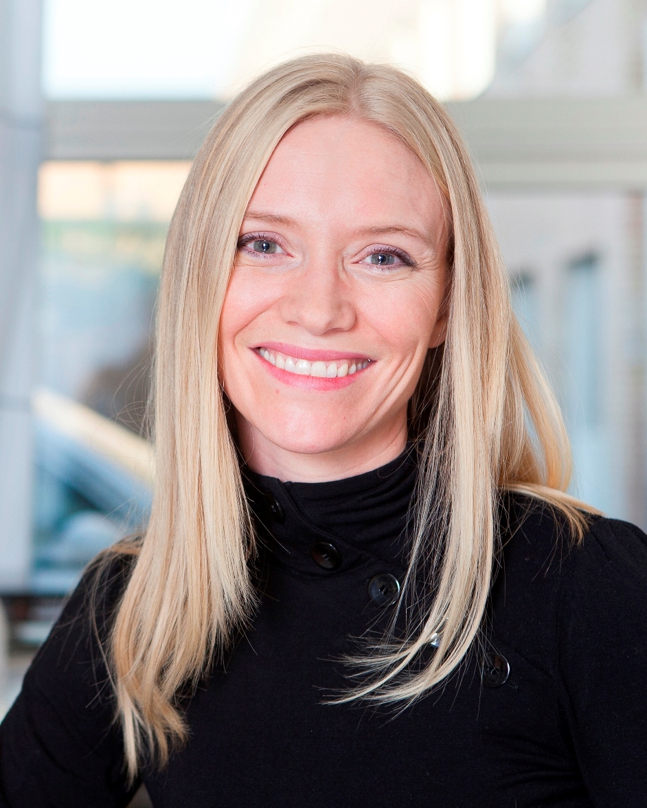
Judith Staerk
In developmental biology a major interest is to identify the molecular mechanisms that direct cell fate decisions and define how stem cells renew and differentiate into more specialized cell types. Pluripotent stem cells can renew indefinitely, while retaining the ability to differentiate into any cell type found in the body. Pluripotency and differentiation are tightly regulated through transcription factor networks, epigenetic mechanisms and metabolism. The Staerk group uses human pluripotent stem cells to understand the interplay between mitochondria, the cells’ metabolism and gene regulation during pluripotent stem cell renewal as well as early blood and neuronal cell specification.
Mitochondria cannot be generated de novo and need to undergo fission and fusion. Proteins involved in the fusion and fission process are the GTPases optic atrophy a (OPA1) and dynmin-like 1 protein (DNM1L). Loss of OPA1 and DNM1L in mice is embryonic lethal due to growth retardation and gross morphological abnormalities. In humans, heterozygous mutations in both OPA1 and DNM1L are associated with autosomal dominant optic atrophy (ADOA). Our group studies whether / how OPA1 and DNM1L deficiency but also overexpression alters the differentiation potential of hESC to CD34+ and blood lineage cells, and how these processes impact on nuclear DNA methylation, transcription and signaling pathways.
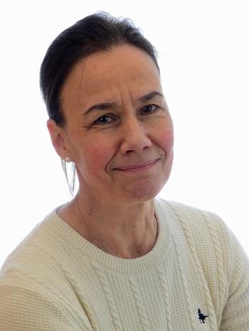
Hilde Nilsen group
We study how DNA repair impact human health in a life course perspective. We study how DNA repair mechanisms protect us from ageing and age-related diseases and use iPSC to model human DNA repair diseases and determine functional consequences in different cell types. We are particularly interested to model motoneurons. We also use patient derived iPCS of the rare DNA repair syndrome Ataxia Telangiectasia to look at genotype-phenotype relationships.
Helge Ræder group
My broad research aim is to understand disease processes leading to pediatric endocrine diseases such as diabetes and disorders of the ovaries and testes. In addition to clinical and genetic studies, we are using cell models based on induced pluripotent stem cells (iPSCs) generated from fibroblasts from patients with endocrine disease. We have mainly focused on making pancreatic beta-like cells, ovarian cells and testicular cells and characterized these cells with omics metods (i.e. proteomics) and functional studies.
Among our research achievements, we have been successful in making human iPSCs from fibroblasts from several members in MODY (Maturity-Onset Diabetes of the Young) families, direct these cells towards pancreatic beta-like cells and characterize their proteome.
Some notable recent papers:
Furuyama, K., S. Chera, L. van Gurp, D. Oropeza, L. Ghila, N. Damond, H. Vethe, J. A. Paulo, A. M. Joosten, T. Berney, D. Bosco, C. Dorrell, M. Grompe, H. Raeder, B. O. Roep, F. Thorel and P. L. Herrera (2019). "Diabetes relief in mice by glucose-sensing insulin-secreting human alpha-cells." Nature 567(7746): 43-48.
Vethe H, Ghila L, Berle M, Hoareau L, Haaland OA, Scholz H, Paulo JA, Chera S, Raeder H: The Effect of Wnt Pathway Modulators on Human iPSC-Derived Pancreatic Beta Cell Maturation. Front Endocrinol (Lausanne) 2019;10:293
Vethe H, Bjorlykke Y, Ghila LM, Paulo JA, Scholz H, Gygi SP, Chera S, Raeder H: Probing the missing mature beta-cell proteomic landscape in differentiating patient iPSC-derived cells. Sci Rep 2017;7:4780 Bjørlykke Y, Søviknes AM, Hoareau L, Vethe H, Mathisen AF, Chera S, Vaudel M, Ghila L and Ræder H. Reprogrammed cells display distinct proteomic signatures associated with colony morphology variability". Stem Cells International, Stem Cells Int 2019;2019:8036035
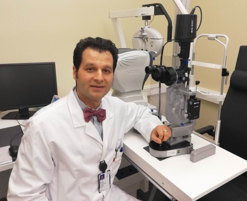
Goran Petrovski group
Goran Petrovski is a professor and senior consultant in vitreoretinal and cataract surgery at the Department of Ophthalmology, Oslo University Hospital and University of Oslo. His hybrid background in medicine, biochemistry and molecular biology and engineering provide a unique environment for research, development and innovation, while his national and international collaborative network play a crucial part of the successes achieved.
Professor Petrovski leads the Center for Eye Research, where basic and translational research involving stem cells meet in one place: 1) cornea limbal- and stromal- stem cell research and transplantation studies, 2) 3D printing of stem cell-laden corneas and femtosecond laser-assisted transplantation techniques, 3) production of iPSC cells from hematopoietic cells and fibroblast, 4) production of iPSC-derived retinal pigment epithelium (RPE) from age-related macular degeneration patients and controls, 5) transplantation of RPE cells on a biocarrier into pigs, 6) cancer stem cell research in uveal melanomas
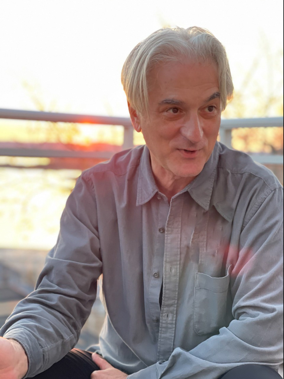
Our translational research focuses on the development of iPS cell-based in vitro neurological disease models. We have reprogrammed iPS cells from patients with amyotrophic lateral sclerosis/frontotemporal dementia spectrum (ALS/FTD: SOD1 and C9orf72 mutations), spinocerebellar ataxia 14 (SCA14: PKCgamma mutation), dystonia (ACTB and GNAL mutations), and an idiopathic familial dysautonomia/peripheral neuropathy syndrome (uncharacterized mutation). We have developed differentiation protocols for neuronal precursors (NPCs), several neuron types (motoneurons, glutamatergic cortical neurons, generic GABAergic neurons) and astrocytes. We have also established a differentiation protocol for microglia developed by the group of Tormod Fladby, and we are working on a differentiation protocol for skeletal muscle and cardiomyocytes. We use these cell types to create 2D and 3D multiple cell type, compartmentalized microfluidic platforms, wherein the principal cell types involved in a disease are positioned in adjacent compartments permitting intercellular communication that simulates the anatomical and physiological situation in vivo. We are also developing a cerebellar organoid platform as a model for SCA14.
This translational research builds on decades of basic research into the structure, function and development of brainstem and spinal cord sensorimotor circuits in animal models, spanning from molecular programs of differentiation to patterns of synaptic connectivity to motor behavior, and on the mechanisms underlying adaptive plasticity following spinal cord injury.
Relevant publications from translational studies:
Sigurjonsson OE, Perreault MC, Egeland T, Glover JC. (2005) Adult human hematopoietic stem cells produce neurons efficiently in the regenerating chicken embryo spinal cord. PNAS 102:5227-5232.
Boulland JL, Halasi G, Kasumacic N, Glover JC (2010) Xenotransplantation of human stem cells into the chicken embryo. J Vis Exp. 2010 Jul 11;(41). pii: 2071. doi: 10.3791/2071.
Boulland JL, Leung DS, Thuen M, Vik-Mo E, Joel M, Perreault MC, Langmoen IA, Haraldseth O, Glover JC (2012) Evaluation of intracellular labeling with micron-sized particles of iron oxide (MPIOs) as a general tool for in vitro and in vivo tracking of human stem and progenitor cells. Cell Transplant. 21(8):1743-1759.
Boulland JL, Mastrangelopoulou M, Boquest AC, Jakobsen R, Noer A, Glover JC, Collas P (2012) Epigenetic Regulation of Nestin Expression During Neurogenic Differentiation of Adipose Tissue Stem Cells. Stem Cells Dev. 22(7):1042-1052.
Boulland JL, Lambert FM, Züchner M, Ström S, Glover JC (2013) A neonatal mouse spinal cord injury model for assessing post-injury adaptive plasticity and human stem cell integration. PLoS One. 8(8):e71701.
Skogseid IM, Røsby O, Konglund A, Connelly JP, Nedregaard B, Jablonski GE, Kvernmo N, Stray-Pedersen A, Glover JC (2018) Dystonia-deafness syndrome caused by ACTB p.Arg183Trp heterozygosity shows striatal dopaminergic dysfunction and response to pallidal stimulation. J Neurodev Disord. 10(1):17.
Liang KX, Vatne GH, Kristiansen CK, Ievglevskyi O, Kondratskaya E, Glover JC, Chen A, Sullivan GJ, Bindoff LA (2021) N-acetylcysteine amide ameliorates mitochondrial dysfunction and reduces oxidative stress in hiPSC-derived dopaminergic neurons with POLG mutation. Exp Neurol. 337:113536.
Akkouh IA, Hughes T, Steen VM, Glover JC, Andreassen OA, Djurovic S, Szabo A. (2021) Transcriptome analysis reveals disparate expression of inflammation-related miRNAs and their gene targets in iPSC-astrocytes from people with schizophrenia. Brain Behav Immun. 94:235-244.
Szabo A, Akkouh IA, Vandenberghe M, Osete JR, Hughes T, Heine V, Smeland OB, Glover JC, Andreassen OA, Djurovic S (2021) A human iPSC-astroglia neurodevelopmental model reveals divergent transcriptomic patterns in schizophrenia. Transl Psychiatry. 11(1):554.
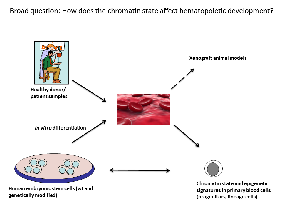
Background: Our research team is generally interested in understanding molecular processes during normal and malignant hematopoiesis. Hematopoiesis describes the formation of all blood cells throughout life. This is ensured by long-term repopulating hematopoietic stem cells (HSC) that give rise to HSC and progenitors with limited renewing capacity. These progenitors produce lineage specific daughter cells that in turn form terminally differentiated erythroid/myeloid and lymphoid cell types. HSC division and blood cell differentiation is tightly regulated by genetics and epigenetic events as well as activation of specific signaling pathways. The dysregulation of one or more of these biological processes can lead to various blood disorders such as leukemia.
Figure 1: Overview of general approaches and methodologies used in my group. The broad research aim of my group is to identify key molecular processes that govern hematopoietic specification, hematopoietic stem cell renewal and differentiation, and differentiation of blood progenitors into mature blood cells. We are using primary patient samples, patient-derived iPSC, genetically modified hESC lines encoding reporter genes to monitor mesoderm and blood differentiation as well as in vivo zebrafish and mouse models.
Research in the Staerk group aims to decipher molecular processes that govern normal and malignant hematopoiesis and focuses on: i) identifying epigenetic and genetic events and signaling pathways that are crucial for human blood development, ii) deciphering the mechanisms by which the nuclear lamina influences hematopoietic development, and iii) identify underlying molecular causes of myeloid blood disorders triggered by defects in lineage differentiation that we study in the physiologic setting. To achieve these goals, we are using human pluripotent stem cells, in vitro differentiation assays as well as animal models along with primary patient samples and we combine these assays with genetic and genomic approaches. Insight obtained from our research projects will help to identify factors needed for efficient blood cell differentiation, and how this differentiation is affected in disease. Further, it will help to identify factors needed to expand functional HSC isolated from bone marrow (BM), and to derive functional HSC from pluripotent stem cells in vitro.
Epigenetic changes during human blood development
One main focus of my group is to understand how epigenetic signatures influence cell fate determination during mesoderm and hematopoietic cell specification. DNA methylation is an epigenetic modification which is key to numerous processes including regulation of gene expression and maintaining genomic integrity. Additional complexity to the overall gene regulation has been added by the discovery of Ten-Eleven-Translocation (TET) enzymes, which are dioxygenases that catalyze the conversion of 5-methylcytosine (5mC) to 5-hydroxymethylcytosine (5hmC). The general importance of 5mC and 5hmC during development has been documented in animal models. Recently, we characterized 5hmC distribution in CD34+ cells and mature blood lineage cells and showed that in CD34+ cells, 5hmC primes expression of genes that regulate myeloid and lymphoid lineage commitment. Throughout blood cell differentiation, intragenic 5hmC is maintained at genes that are highly expressed and required for acquisition of the mature blood cell phenotype. Moreover, in CD34+ cells, 5hmC at enhancers was associated with increased binding of RUNX1 and FLI1 that are TFs crucial for hematopoiesis. To further investigate the role of 5mC and 5hmC during human blood cell differentiation, we now established oxidative bisulfite sequencing and deleted endogenous DNMT3A and 3B or different TET family members in hESC lines encoding reporter genes that allow monitoring blood cell differentiation. We aim to decipher how epigenetic changes and transcriptional networks interact during blood cell formation, and how 5mC as well as 5hmC influence the recruitment of key TFs during human hematopoiesis. Critical to this question is how epigenetic modifiers such as DNMT and TET enzymes interact with non-coding-RNAs (nc-RNAs) and which nc-RNA networks are required to generate mesoderm and hematopoietic progenitor cells from hESC.
Role of the nuclear lamina during human hematopoiesis
More recently, the group developed an interest in the nuclear lamina (NL)/lamin proteins. Lamins are divided into A-type lamins that are expressed in most somatic cell types, but are not expressed in stem cells, while B-type lamins (LMNB1 and LMNB2) are highly expressed in both, stem cells and differentiated cell types. Apart from the well-established role in forming a scaffold underneath the inner nuclear membrane, the NL has been implicated in nuclear positioning of chromatin and transcriptional regulation, which is thought to be critical for cell fate decisions. We aim to address how lamin-mediated gene regulation influences human hematopoiesis, and whether lamin proteins are dysregulated in blood disorders. Lamin proteins tether chromatin to the NE and influence transcription, signaling networks and cell fate decisions. Deciphering lamin-mediated gene regulation during blood cell specification will increase our understanding of underlying regulatory mechanisms during blood cell differentiation in the physiologic setting and will help to elucidate whether these processes are affected in disease.
Institute of Medical Microbiology, Section for Molecular Biology, is localised at Oslo University Hospital, Rikshospitalet, Gaustad, Oslo. Four different research groups are colocalised, concentrating on DNA-repair and epigenetic studies, brain development, microbiology and biocomputing.
We have a very close collaboration with the Bjørås group which is also a part of the Stem Cell Centre, as well as biocomputing collaborations with the Rognes group. For characterization of knockout mice we collaborate with the Falnes (University of Oslo) and Krokan (University in Trondheim) groups. We also collaborate with excellent research groups in Germany, France, China, Holland, UK and USA.
Our aim is to elucidate new mechanisms of demethylation and hydroxylation required for for repair and regulation of the mammalian (epi)genome and to address (epi)genome instabilities associated with diseases such as cancer. Our current models also include genes affecting post translational modifications in RNA and proteins.
Currently we have MSc ("hovedfag"), PhD students and post docs from The Agricultural University, University of Oslo, University of Trondheim ("Siv. Ing."), Austria, Sweden, The Phillipines, Germany and USA.
Selected publications (2006-2012):
Robertson AR, Dahl JA, Ougland R and Klungland A (2012) 5-hydroxymethylcytosine DNA pull down using JBP1-coated magnetic beads. Nature protocols, 7:340-50
Robertson A, Dahl JA, Vågbø C, Tripathi P, Krokan H and Klungland A (2011) A novel method for the efficient and selective identification of 5-hydroxymethylcytosine in genomic DNA. Nucleic Acids Res39:e55*
*The method described is patented and released by ZYMO research, USA.
van den Born E*1, Vågbø CB*1, Songe-Møller L*1, Leihne V, Lien GF, Krokan HE, Kirpekar F, Klungland A*2 and Falnes PØ*2 (2011) ALKBH8 mediated hydroxylation of a novel diastereomeric pair of wobble nucleosides in tRNA. Nature Commun 2, 172 *1Contributed equally *2 Corresponding
Haj-Yasein NN, Vindedal GF, Eilert-Olsen M, Gundersen GA, Skare Ø, Laake P, Klungland A, Thorén AE, Burkhardt JM, Ottersen OP and Nagelhus EA (2011) Glial-conditional deletion of aquaporin-4 (Aqp4) reduces blood-brain water uptake and confers barrier function on perivascular astrocyte endfeet. Proc Natl Acad SciUSA 108:17815-20
Thrane AS*, Rappold PM*, Fujita T, Torres A, Bekar LK, Takano T, Peng W, Wang F, Thrane VR, Enger R, Haj-Yasein NN, Skare Ø, Holen T, Klungland A, Ottersen OP, Nedergaard M and Nagelhus EA (2011) Critical role of aquaporin-4 (AQP4) in astrocytic Ca2+ signaling events elicited by cerebral edema.Proc Natl Acad Sci USA108:846-51 *Contributed equally
Strømme S, Dobrenis K, Sillitoe R, Gullinello M, Ali N, Davidson C, Micsenyi MC, Stephney G, Ellevog L, Klungland A and Walkley SU (2011) Sodium/Hydrogen exchanger related mental retardation syndrome: evidence for endosomal-lysosomal dysfunction in Slc9a6 targeted mice. Brain
Møllersen L*, Rowe AD*, Larsen E, Rognes T and Klungland A (2010) Two modes of somatic CAG repeat expansions in Huntington disease transgenic mice. PLoS Genet 9;6 *Corresponding
Songe-Møller L*1, van den Born E*1, Leihne V*1, Vågbø CB*1, Kristoffersen T, Krokan HE, Kirpekar F, Falnes PØ*1,2 and Klungland A*1,2 (2010) Mammalian ALKBH8 possesses tRNA methyltransferase activity required for the biogenesis of multiple wobble uridine modifications implicated in translational decoding. Mol Cell Biol 30:1814-27 *1Contributed equally *2Corresponding
Lauritzen KH, Moldestad O, Eide L, Carlsen H, Nesse G, Storm JF, Mansuy IM, Bergersen LH* and Klungland A* (2010) Mitochondrial DNA toxicity in forebrain neurons causes apoptosis, neurodegeneration, and impaired behavior. Mol Cell Biol 30:1357-67 *Corresponding
Saxowsky T, Meadows K, Klungland A and Doetsch P (2008) 8-Oxoguanine-mediated transcriptional mutagenesis causes Ras activation in mammalian cells. Proc Natl Acad Sci USA 105:18877-82
Larsen E*, Kleppa L*, Meza TJ, Meza-Zepeda LA, Rada C, Castellanos CG, Lien GF, Nesse GJ, Neuberger MS, Lærdahl JG, Doughty RW and Klungland A (2008) Early onset lymphoma and extensive embryonic apoptosis in two domain-specific Fen1 mice mutants. Cancer Res 68:4571-9 *Contributed equally
Ringvoll J, Nabong MP, Nordstrand LM, Meira L, Pang B, Dedon P, Bjelland S, Samson LD, Falnes PØ and Klungland A (2008) AlkB homolog 2 (ABH2) is the principal mammalian dioxygenase for repair of ethenoadenine lesions in DNA. Cancer Res 68:4142-9
Kovtun IV, Liu Y, Bjørås M, Klungland A, Wilson SH and McMurray CT (2007) OGG1 initiates age-dependent CAG trinucleotide expansion in somatic cells. Nature447:447-52
Leiros I*, Nabong MP*, Grosvik K, Ringvoll J, Haugland GT, Uldal L, Reite K, Olsbu IK, Knaevelsrud I, Moe E, Andersen OA, Birkeland NK, Ruoff P, Klungland A and Bjelland S (2007) Structural basis for enzymatic excision of N1-methyladenine and N3-methylcytosine from DNA. EMBO J 26:2206-17 *Contributed equally
Ringvoll J, Nordstrand LM, Vågbø CB, Talstad V, Reite K, Aas PA, Lauritzen KH, Liabakk NB, Bjørk A, Doughty RW, Falnes PØ, Krokan HE, Klungland A (2006) Repair deficient mice reveal mABH2 as the primary oxidative demethylase for repairing 1meA and 3meC lesions in DNA. EMBO J 25:2189-98