
Services
| The facility includes three confocal microscopes and three whole-cell fluorescence setups as well as software for image enhancement. The confocal microscopes are located in rooms 4034 and 4035. |
|
This inverted point-scanning confocal microscope with high spatial resolution is well-suited for static imaging. Common applications include high resolution immuno-fluorescence for studying protein localization (pictured below), and colocalization using traditional antibody labelling as well as Duolink. The microscope is fitted with an incubation chamber (Zeiss XL S1) for live-cell imaging experiments requiring control of temperature and O2/CO2 levels. The microscope is also currently being configured for FRET. Laser lines: 458, 488, 514, 543, 633 nm. |
|
This inverted line-scanning confocal microscope is well-suited for imaging rapid events such as Ca2+ sparks. The 7 Live provides high-speed scanning, with acquisition rates of 1000 images of 512 x 512 pixels in less than 10 seconds. The microscope is set up with a perfusionsystem (37°C), patch clamp equipment (Axon Axopatch 200B) and field stimulation. Flash photolysis equipment is also available. Laser lines: 488, 532 nm. |
|
This inverted point-scanning confocal microscope has modest spatial and temporal resolution, and is commonly employed for both imaging of ionic fluorophores and static imaging protein localization / colocalization. This setup is also equipped with whole-cell fluorescence with PMT-based detection (Ratio Master, Photon Technology International, pictured at right), a heated perfusion system, electrophysiology (Axon Axoclamp 2B) and field stimulation. Laser lines: 488, 543, 633 nm. |
| In conjunction with the confocal core facility, three setups for whole-cell fluorescence are available by collaboration with Core Facility staff: |
|
This fluorescence system from Photon Technology International enables ratiometric wide-field fluorescence detection (Easy Ratio Pro)by high-speed camera (Orca Flash 4.0, Hamamatsu Photonics) with WarpDrive interface controller (pictured right). The setup is also equipped with a CellTester system (World Precision Instruments), enabling controlled stretch and force measurement of individual cardiomyocytes. The setup contains a heated perfusion system, as well as equipment for electrophysiology (Axon Axoclamp 200B) and field stimulation. |
|
Cairn Optoscan Wide-field Fluorescence / Zeiss Axiovert 200 This inverted wide-field microscope is employed for studying ionic dynamics in live cells. Whole-cell fluorescence can be measured using a fluorescence system from Cairn Research (Optoscource / Optoscan, 1 detection channel). The setup is equipped with a heated perfusion system, as well as equipment for measuring cell shortening (Crescent electronics) and electrophysiology (Axon Axoclamp 2B) and for exciting cells by field stimulation. |
|
Photon Technology International Wide-Field Fluorescence / Zeiss Axio Observer This inverted wide-field microscope is employed for studying ionic dynamics in live cells. Whole-cell ratiometric fluorescence (for example Fluo-4, Fura-2, SBFI) can be measured using the Ratio Master system from Photon Technology International employing Felix software. The setup is equipped with a heated perfusion system, equipment for measuring cell shortening (Crescent electronics) and electrophysiology (Axon Axoclamp 2A) and for exciting cells by field stimulation. |
|
MMI Cellcut Laser Dissection Microscope / Nikon Eclipse TE 2000-S The Cellcut system enables rapid, precise isolation of cells and tissue from a variety of sample types including fresh, frozen, or embedded tissues and cells. Regions of interest are marked and cut automatically using a precisely focused UV-Laser for further analyses. |
|
Huygens deconvolution software from Scientific Volume Imaging The core facility is licensed for use of deconvolution software from SVI which improves image quality by reversing optical distortion in the image. |
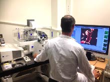 Zeiss LSM 710 Confocal / Axio Observer
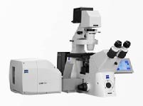 Zeiss LSM 7 Live Confocal / Axio Observer
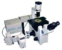 Zeiss LSM 510 Confocal / Axiovert 100
 PTI Easy Ratio Pro Wide-field Fluorescence / Zeiss Observer D1